Home » Diseases » Emergencies / First Aid for Dogs & Cats – General Information » Emergencies / First Aid A-Z » Gastric Dilatation-Volvulus (GDV)
Gastric Dilatation-Volvulus (GDV)
Gastric dilatation-volvulus (GDV) is an acute, life-threatening condition that primarily affects medium and large breed dogs. In this condition, the stomach rotates around its own longitudinal axis, causing both the stomach entrance (cardia) and exit (pylorus) to close. This rotation creates a closed system in which gases and fluids accumulate, leading to massive gastric distension. The rotation can range from 90° to 360°, with a rotation of 180° to 270° being most common.
Anatomically, the dog’s stomach is only attached to the spleen, liver, and diaphragm by small ligaments (ligamenta), allowing for some mobility. In gastric dilatation-volvulus, the stomach usually shifts clockwise when viewed from behind the dog. The pylorus moves from the right side of the abdomen to the left and dorsally (towards the back), while the greater curvature of the stomach moves from left ventral (towards the belly) to the right.
The pathophysiological consequences are severe: The rotation compresses blood vessels, leading to circulatory disturbances in the stomach wall. The resulting ischemia can cause necrosis (tissue death) of the stomach wall. Additionally, the massive distension of the stomach puts pressure on the diaphragm, impairing breathing and hindering venous return to the heart, which can lead to hypovolemic shock.
Causes
In a gastric dilatation-volvulus, the stomach rotates around its longitudinal axis from its usual position, preventing air or other stomach contents from escaping. This severely restricts blood supply to the stomach and partially to the spleen. The accumulation of gases in the stomach causes a barrel-shaped enlarged stomach. The poor blood circulation and increased pressure in the stomach lead to the death of the affected tissue.
Mostly large dog breeds are affected, such as Great Danes and German Shepherds. But breeds like Akita Inu and Shar-Pei also seem particularly susceptible to experiencing GDV. Feeding dry food and subsequent intake of large amounts of fluid can also contribute to GDV (Fig.). For these dog breeds and dogs that have already experienced GDV, smaller meals should be fed more frequently throughout the day.
If surgery takes place within 6 hours after the occurrence of GDV, the prospects for success are good. The prognosis for successful therapy is 80-90%. The more time that passes between suffering from GDV and the predominantly surgical restoration of normal anatomical conditions, the worse the chances of success.
Supplement
The exact etiology of GDV is multifactorial and not fully understood to this day. However, there are several predisposing factors that can increase the risk of GDV.
The anatomical risk factors primarily include the deep, narrow chest of certain dog breeds such as Great Danes, German Shepherds, Dobermans, Irish Wolfhounds, Bernese Mountain Dogs, Weimaraners, and Akita Inus. This anatomy provides the stomach with more room for movement in the abdominal cavity. A loose gastric ligament (Ligamentum hepatogastricum) can also contribute to its development.
Nutritional factors also play a significant role. Feeding large amounts of food once a day significantly increases the risk as it greatly stretches the stomach. Dry food that swells in the stomach, especially when followed by the intake of large amounts of water, presents another risk factor. Rapid eating with air swallowing (aerophagia) promotes gas accumulation in the stomach.
Behavioral factors include intense physical activity immediately after feeding, which can delay gastric emptying and promote gas formation. Stress can also contribute to gas accumulation through increased air intake during panting.
Genetic factors are also discussed, as GDV occurs more frequently in some families. A familial predisposition has been demonstrated in several studies, suggesting a hereditary component.
Age and gender also influence the risk: Older dogs (>7 years) are more frequently affected, presumably due to decreasing tissue tension. In some breeds, male animals seem to have a higher risk, although the exact mechanism for this is still unclear.
Symptoms
- Restlessness
- Increasing, sometimes massive gas accumulation in the stomach 1 to 2 hours after the last feeding, associated with a large abdominal circumference
- Unsuccessful attempts to vomit
- Shock symptoms
The clinical signs of GDV typically develop rapidly and are characterized by their dramatic nature. The symptoms are often observed within 1-2 hours after the last feeding, but can also occur at other times.
The most noticeable symptom is the progressive gaseous distension of the abdomen, leading to a clearly visible, barrel-shaped swelling of the belly. This distension is particularly pronounced in the area of the front half of the abdomen and asymmetrical, with the left side often more affected. Upon palpation, the abdomen feels tense and drum-like.
Affected dogs show increasing restlessness, manifesting as restless pacing, whining, or a characteristic posture with an arched back and lowered head. Often, the animals try to vomit, but due to the gastric obstruction, this remains unsuccessful (unproductive retching). This symptom is particularly valuable diagnostically.
As the condition progresses, signs of hypovolemic shock develop: The mucous membranes become pale to cyanotic (bluish), the capillary refill time extends to over 2 seconds. The pulse becomes rapid and thready, while the respiratory rate increases due to the pressure of the bloated stomach on the diaphragm. The body temperature may initially be elevated but decreases in advanced stages of shock.
The dogs show increasing weakness and apathy, may express pain when palpating the abdomen, and often assume a prayer position to reduce pressure in the abdominal cavity. In advanced stages, collapse, impaired consciousness, and eventually death occur if immediate veterinary intervention is not provided.
- In some dogs, cardiac arrhythmias may additionally occur, caused by the release of myocardial toxins from the ischemic gastric tissue and by electrolyte shifts. These arrhythmias can occur even 24-72 hours after initial treatment and represent a dangerous complication.
First Aid
- Gastric dilatation-volvulus is one of the most severe emergencies in small animal medicine.
- Immediate (!) transport to a veterinary hospital is necessary. Without professional help, this emergency cannot be resolved and will lead to the animal’s death within a few hours. Surgery is usually necessary.
- Do not attempt to remove the gas from the stomach yourself.
- Do not administer anything orally.
- Watch for early symptoms such as drooling, unsuccessful attempts to vomit, restlessness, expressions of pain, and difficulty breathing.
Recurrence rates after gastropexy are low. High preoperative lactate levels, gastric wall necrosis, necessary splenectomy, existing peritonitis or sepsis are considered prognostically unfavorable factors. In the postoperative phase, cardiac activity should be monitored and shock treatment continued.
Diagnosis
The diagnosis of gastric dilatation-volvulus is made through a combination of anamnesis, clinical examination, and imaging procedures. Due to the urgency of the condition, the diagnosis must be made quickly and precisely.
In the anamnesis, information about breed, age, feeding habits, and current symptoms is crucial. The clinical examination typically shows a bloated abdomen with a tympanic sound on percussion. Auscultation of the abdomen reveals reduced or absent intestinal sounds. Vital signs indicate a state of shock: tachycardia, weak pulse, prolonged capillary refill time, and pale mucous membranes.
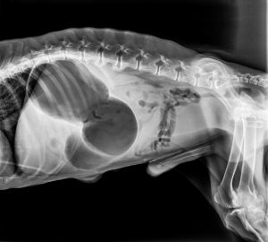
Imaging diagnostics are essential for confirming the diagnosis. X-rays of the abdomen in right lateral recumbency are the method of choice. The characteristic X-ray image shows a severely gas-filled, compartmentalized stomach with a typical “double bubble” or “popcorn sign”, created by the displacement of the pylorus. The spleen may also appear displaced. In unclear cases, contrast medium administration or ultrasound examination can help differentiate between simple gastric distension and gastric dilatation-volvulus.
Various laboratory parameters are relevant: An elevated hematocrit indicates dehydration, while electrolyte shifts (especially hypokalemia) and metabolic acidosis indicate shock. Blood lactate concentration is an important prognostic parameter – values above 6 mmol/l are associated with increased mortality and indicate severe tissue damage. Elevated liver enzymes and kidney parameters may indicate organ damage due to shock.
An ECG should be performed to detect cardiac arrhythmias that may occur due to electrolyte shifts or myocardial damage. Ventricular arrhythmias are common complications and can occur even days after initial treatment.
Differential diagnoses must exclude other causes of acute abdominal pain and bloating, such as simple gastric distension, foreign body ileus, acute pancreatitis, or intestinal volvulus.
Further veterinary measures
In the rarest cases, therapy without surgery is possible.
Therapy (shock treatment and surgery) is initiated immediately upon the animal’s arrival at the hospital.
The prognosis is poor if:
- the torsion has existed for >6 hours,
- high lactate levels are present in the blood,
- the gastric wall has already partially died and must be removed (partial gastric resection or invaginating suture),
- the spleen must also be removed,
- peritonitis and/or sepsis are already present.
At the end of the surgery, fixation of the stomach to the abdominal wall using special methods (gastropexy) is recommended. Gastropexy prevents the stomach from relocating again. The recurrence rate, meaning the reoccurrence of gastric dilatation-volvulus at a later time, is significantly reduced.
Supplements
The treatment of gastric dilatation-volvulus is a veterinary emergency that requires rapid and coordinated action. The therapy includes several phases: initial stabilization, decompression of the stomach, and surgical intervention.
Stabilization begins with aggressive fluid therapy to treat hypovolemic shock. Crystalloid solutions (e.g., Ringer’s lactate) are administered at high doses (90 ml/kg/h) as an initial shock dose, followed by a maintenance dose. In severe shock conditions, colloids or plasma expanders may also be used. The electrolyte balance, especially potassium levels, must be monitored and corrected.
Decompression of the stomach is the next critical step. For this, an orogastric tube is inserted under light sedation to drain gases and fluids. If tube insertion is not possible due to the gastric torsion, percutaneous trocarization of the stomach may be considered – a procedure that, however, is associated with risks such as peritonitis and should only be performed by experienced veterinarians.
The definitive therapy is surgical and should be performed as quickly as possible after initial stabilization. After median laparotomy, the stomach is manually rotated back into its physiological position (usually counterclockwise). Then, a thorough inspection of the gastric wall for necrosis or perforations follows. Damaged tissue must be resected. The spleen is also examined, as it is often affected by the rotation and may require splenectomy.
To prevent recurrence, a gastropexy is performed – the permanent fixation of the stomach to the abdominal wall. Various techniques are available:
- Incisional gastropexy: An incision in the gastric wall is sutured to an incision in the right abdominal wall
- Belt-loop gastropexy: A strip of the gastric wall is pulled through a tunnel in the abdominal wall and fixed
- Laparoscopic gastropexy: Minimally invasive technique with lower morbidity
Intensive monitoring is required postoperatively. Fluid therapy, analgesia, and antibiotic administration are continued. ECG monitoring is particularly important, as ventricular arrhythmias can occur 24-72 hours after surgery and may need to be treated with antiarrhythmic drugs (e.g., lidocaine). Feeding is gradually resumed, starting with small amounts of water about 12-24 hours postoperatively, followed by easily digestible food in small, frequent portions.
Sepsis and shock treatment are continued after surgery.
The greatest risk after surgery (24-36 hours) is cardiac arrhythmias. Electrocardiographic monitoring is necessary and has a good prognosis with timely, sometimes repeated administration of appropriate medications.
Prognosis and aftercare
The prognosis for gastric dilatation-volvulus depends significantly on the timing of intervention and the extent of tissue damage already present. With early diagnosis and adequate treatment, the survival rate is between 80 and 90%. However, mortality increases significantly if gastric wall necrosis, peritonitis, or advanced shock are already present.
Prognostically unfavorable factors include:
- Delay between symptom onset and surgical intervention of more than 6 hours
- Preoperative lactate levels >6 mmol/l that do not decrease after initial fluid therapy
- Necessity for partial gastrectomy due to necrosis
- Required splenectomy
- Existing peritonitis or sepsis
- Severe cardiac arrhythmias
Postoperative care begins immediately after surgery with intensive monitoring of vital parameters, particularly heart function. Continuous ECG monitoring for 48-72 hours is recommended, as ventricular arrhythmias are a common and potentially life-threatening complication. Fluid and electrolyte therapy is continued until the patient independently takes in sufficient water.
Wound inspection is performed daily to detect infections or dehiscence early. Analgesia is continued for at least 3-5 days to control pain and reduce stress. Antibiotics are administered for 5-10 days depending on intraoperative findings.
Nutrition is gradually reintroduced: After 12-24 hours, small amounts of water can be offered, followed by easily digestible food in small, frequent portions after 24-48 hours. In the long term, feeding should be changed to 3-4 smaller meals daily. The use of elevated food bowls is controversial – recent studies suggest that they might even increase the risk of recurrent gastric dilatation volvulus.
Physical activity should be restricted for 10-14 days to ensure optimal wound healing. After that, exercise can be slowly increased, but intense activity immediately after feeding should be permanently avoided.
Regular follow-up examinations are important to monitor the healing process and detect potential complications early. The first check-up usually takes place 10-14 days postoperatively to remove skin sutures, with further checks after 1, 3, and 6 months.
Summary
Gastric dilatation volvulus (GDV) is an acute, life-threatening condition that primarily affects large and deep-chested dog breeds. In this condition, the stomach rotates around its own longitudinal axis, blocking both entrance and exit, causing gases and fluids to accumulate in the stomach. This leads to massive distension of the stomach with subsequent impairment of blood circulation and compression of adjacent organs.
The etiology is multifactorial and includes anatomical, nutritional, behavioral, and genetic factors. The main risk factors include large, deep-chested breeds, feeding large amounts once daily, rapid eating with air swallowing, and intense exercise after feeding.
Clinical symptoms develop rapidly and include a bloated abdomen, unproductive retching, restlessness, salivation, and increasing signs of shock. Diagnosis is based on history, clinical examination, and imaging techniques, with radiographs being the most important diagnostic tool.
Treatment must be immediate and includes shock management, gastric decompression, and surgical intervention with repositioning of the stomach and gastropexy to prevent recurrence. Postoperative monitoring focuses on cardiac arrhythmias, wound healing, and gradual resumption of feeding.
The prognosis is good with early intervention but worsens with increasing duration of the condition and the occurrence of complications such as gastric wall necrosis or peritonitis. Preventive measures include several small meals daily, avoiding intense activity after feeding, and possibly prophylactic gastropexy in predisposed breeds.
Despite improved treatment options, gastric dilatation volvulus remains one of the most dangerous emergency situations in small animal medicine and requires immediate veterinary action to save the affected animal’s life.
Outlook on current research
Current research on gastric dilatation volvulus in dogs focuses on several promising areas that could improve the understanding, prevention, and treatment of this life-threatening condition.
In the field of genetics, molecular markers associated with an increased risk for gastric dilatation volvulus are increasingly being identified. Studies in predisposed breeds such as Great Danes and Irish Wolfhounds have already identified initial genes that could be linked to anatomical peculiarities of the gastrointestinal tract or connective tissue structure. These genetic investigations could lead to future screening tests that identify particularly vulnerable individuals and enable targeted preventive measures.
Minimally invasive surgical techniques are continuously being developed. Laparoscopic gastropexy as a prophylactic measure in high-risk patients is gaining increasing importance. Recent studies show that this technique is associated with lower postoperative morbidity, shorter hospital stays, and faster recovery, while offering comparable effectiveness to traditional open procedures. Innovative endoscopically assisted techniques that do not require complete laparotomy are in clinical trials.
Biomarker research focuses on identifying reliable prognostic indicators. In addition to the established lactate value, new biomarkers such as troponin I (as an indicator of myocardial damage), C-reactive protein, and specific microRNAs are being investigated, which can provide early indications of tissue damage and organ failure. These could enable more precise risk prediction and individualized treatment strategies.
In the field of intensive care, new protocols for shock treatment are being evaluated. Goal-directed fluid therapy, based on dynamic parameters such as stroke volume variation and systemic vascular resistance, could improve survival rates. The use of vasopressors and inotropes is also being systematically investigated to establish optimal treatment protocols.
Microbiome research has shown that changes in the gut flora can be associated with gastrointestinal motility disorders. Initial studies suggest that certain dysbioses could influence the risk of gastric dilatation volvulus. In the future, probiotic interventions or targeted microbiome modulations could represent preventive approaches.
Nutritional studies are investigating the influence of different food types, feeding frequencies, and nutrient compositions on gastric emptying and gas formation. Special diets that reduce gas formation and optimize gastric emptying could be developed for high-risk patients.
These diverse research approaches promise to significantly improve the management of gastric dilatation volvulus in the coming years – from prevention to acute treatment and aftercare.
Frequently asked questions (FAQs)
- Which dog breeds are particularly at risk of suffering from gastric dilatation-volvulus?
Particularly at risk are large and deep-chested breeds such as Great Danes, German Shepherds, Dobermans, Bernese Mountain Dogs, Irish Wolfhounds, Weimaraners, Akita Inus, and Shar-Peis. Large mixed breeds with a similar body structure can also be affected. - How can I as a dog owner prevent gastric dilatation-volvulus?
Preventive measures include feeding several smaller meals instead of one large one, avoiding intense exercise immediately before and after eating, using special slow-feeder bowls to slow down food intake, and for high-risk patients, possibly a prophylactic gastropexy. - Is gastric dilatation-volvulus always an emergency that requires immediate veterinary assistance?
Yes, gastric torsion is always an acute emergency that can lead to death within a few hours. If gastric torsion is suspected, a veterinarian or veterinary hospital should be consulted immediately, regardless of the time of day or day of the week. - Can gastric dilatation-volvulus recur after successful treatment?
Without surgical fixation of the stomach (gastropexy), the recurrence rate is up to 80%. After a correctly performed gastropexy, the risk drops to below 5%, which is why this operation is routinely performed during surgical treatment. - How long does recovery take after surgery for gastric dilatation-volvulus?
The acute recovery phase lasts about 10-14 days, during which rest and limited movement are important. Full recovery can take 4-6 weeks depending on the severity of the disease and any complications. Regular follow-up checks are important. - What role does food bowl height play in preventing gastric dilatation-volvulus?
The recommendation to offer food in raised bowls is controversial. Recent studies suggest that raised bowls might even increase the risk of gastric dilatation-volvulus in some dogs. Individual consultation with a veterinarian is recommended. - Is a prophylactic gastropexy advisable for my dog?
For dogs of predisposed breeds, especially if they have additional risk factors or relatives who have already been affected by gastric dilatation-volvulus, a prophylactic gastropexy may be advisable. The optimal time is often during an already planned surgery such as neutering. The decision should be made individually with the veterinarian. - What symptoms distinguish simple gastric overload from gastric dilatation-volvulus?
With simple gastric overload, the dog can usually vomit successfully and shows less dramatic symptoms. With gastric dilatation-volvulus, unproductive retching, rapid deterioration of general condition, and a hard, tense, asymmetrically bloated abdomen are characteristic. In case of doubt, a veterinarian should always be consulted. - How high are the treatment costs for gastric dilatation-volvulus?
The costs for emergency care, surgery, and inpatient stay for gastric dilatation-volvulus can range between €1,500 and €4,000 depending on the region and individual complications. Pet health insurance can cover these financial risks. - Can stress trigger gastric dilatation-volvulus?
Stress can lead to increased air swallowing due to increased panting, thereby increasing the risk of gastric dilatation-volvulus. A higher incidence has been observed particularly in situations such as stays in animal shelters, travel, or during weather changes. Stress reduction can therefore be considered a preventive measure.
Literature
- Löwe, G. and Löwe, O. (2021). Emergencies in Dogs and Cats – A Veterinary Guide. Kynos-Verlag. 208 p.
- Gazzola KM, Nelson LL. The relationship between gastrointestinal motility disorders and gastric dilatation-volvulus in dogs. Topics in Companion Animal Medicine. 2021;42:100503.
- O’Neill DG, Case J, Boag AK, et al. Gastric dilation-volvulus in dogs attending UK emergency-care veterinary practices: prevalence, risk factors and survival. Journal of Small Animal Practice. 2017;58(11):629-638.
- Maki LC, Males KN, Byrnes MJ, et al. Incidence of gastric dilatation-volvulus following a prophylactic gastropexy in 782 dogs. Journal of the American Veterinary Medical Association. 2021;258(11):1201-1205.
- Przywara JF, Abel SB, Peacock JT, et al. Occurrence and recurrence of gastric dilatation with or without volvulus after incisional gastropexy. Canadian Veterinary Journal. 2020;61(2):183-188.