Home » Diseases » Emergencies / First Aid for Dogs & Cats – General Information » Emergencies / First Aid A-Z » Traumatic Brain Injury
Traumatic Brain Injury
Traumatic Brain Injury (TBI) represents an acute injury to the brain caused by external force to the skull. In dogs and cats, we distinguish between primary and secondary brain damage. Primary brain damage occurs immediately at the time of trauma and includes direct tissue damage such as contusions, hemorrhages, or axonal injuries. Secondary brain damage develops in the hours and days following the trauma and results from pathophysiological processes such as brain swelling (edema), inflammatory responses, circulatory disturbances, and metabolic changes. These secondary processes can significantly amplify the overall damage and are the main target for therapeutic interventions.
The severity of a traumatic brain injury in veterinary medicine is assessed using a modified Glasgow Coma Scale, which takes into account the animal’s state of consciousness, motor abilities, and brainstem reflexes. This classification is crucial for prognosis and choice of therapy.
Causes
Even minor injuries in the head area can bleed heavily. However, they are usually harmless and heal quickly.
This results in a skull contusion without involvement of the brain.
Nevertheless, as a precaution, allow your pet some rest for the next few hours to days.
However, in case of serious injuries, for example due to a traffic accident or a fall resulting in severe traumatic brain injury, it is an emergency.
In addition to the injury to the skull, there is involvement of the brain, such as brain swelling or bleeding in the brain.
The possible consequences range from a mild concussion to the death of the animal.
The symptoms may only become apparent several hours after the trauma (Fig.).
Supplements
Traumatic brain injuries in pets primarily occur due to traffic accidents, which account for about 60% of cases. Other common causes include falls from great heights (especially in cats), kicks or blows, bite injuries from other animals, and gunshot wounds. For small dog breeds and cats, even a fall from a low height can lead to significant injuries. Brachycephalic breeds such as Pugs, French Bulldogs, or Persian cats have an increased risk of serious consequences from head injuries due to their skull anatomy, as the altered skull shape provides less protection for the brain.
The injury mechanisms can be categorized into acceleration/deceleration traumas (sudden acceleration or deceleration of the head), compression traumas (direct force applied to the skull), and penetration traumas (penetrating objects). Each of these mechanisms leads to different injury patterns in the brain tissue.
Symptoms
- Dizziness
- Head shaking
- Staggering
- Disorientation
- Unconsciousness
- Heavy bleeding
- Bleeding from the nose or ears
- Bruising around the eyes
- Refusal to eat
- Vomiting
- Seizures
- Coma
The clinical signs of traumatic brain injury vary greatly depending on the location and severity of the injury. The most common neurological symptoms include altered consciousness, ranging from mild drowsiness to deep coma. Many animals show coordination disorders such as staggering, circling, or inability to stand. Seizures also occur in about 30% of patients with severe TBI.
Changes in pupil size and reaction are important diagnostic indicators: unequal pupils (anisocoria) or lack of light reaction may indicate increased intracranial pressure or brainstem damage. Other signs include abnormal eye movements (nystagmus), altered body posture and reflexes, as well as respiratory disturbances.
Systemic symptoms include vomiting, cardiac arrhythmias, blood pressure changes, and temperature regulation disorders. Externally visible injuries such as bleeding from the nose or ears, bruising around the eyes, or open skull fractures may indicate a TBI, but are not always present.
First Aid
- Keep your pet as calm as possible
- Do not move it unnecessarily
- Especially do not move the head and neck area
- Carefully remove the collar
- Cover the source of bleeding with a clean cloth
- Do not apply pressure (possible skull fracture?)
- Keep your pet warm
- Take your pet to a veterinary hospital immediately.
- If possible, call ahead to the veterinary hospital.
Diagnosis
The diagnosis of a traumatic brain injury begins with a thorough neurological examination that assesses the state of consciousness, cranial nerve reflexes, motor functions, and posture. The modified Glasgow Coma Scale for animals allows for a standardized assessment of neurological function and classification of severity.
Imaging techniques are essential for precise diagnosis. Computed tomography (CT) is the method of choice in the acute phase, as it can be performed quickly and reliably shows skull fractures, acute bleeding, and major tissue damage. Magnetic resonance imaging (MRI) provides a more detailed view of brain tissue and is particularly valuable for assessing edema, diffuse axonal injuries, and smaller lesions, but requires longer examination time and is not universally available.
Laboratory tests are used to detect systemic complications and include a complete blood count, electrolytes, blood gases, and coagulation parameters. Biomarkers for brain damage such as S100B protein or neuron-specific enolase are increasingly being researched in veterinary medicine but are not yet routinely used.
Further veterinary measures
In case of a serious traumatic brain injury, a computed tomography scan of the head is the most important measure.
Other injuries, such as an injury to the cervical spine, must be ruled out radiologically and the patient should be kept as still as possible until then.
Depending on the results of the radiological examinations, surgeries may be necessary.
For patients with severe traumatic brain injury, heart activity, blood pressure, respiration, and body temperature, as well as water and electrolyte balance, must be monitored and therapeutically supported if necessary.
Usually, infusion therapy and sometimes artificial nutrition are provided.
Supplements
The treatment of traumatic brain injury primarily aims to minimize secondary brain damage and ensure basic brain supply. Stabilizing vital functions is of highest priority: securing the airways, supporting respiration, and maintaining adequate blood pressure are crucial for cerebral perfusion.
The medicinal therapy encompasses several approaches. To control brain edema, osmotically active substances such as mannitol (0.5-1.5 g/kg i.v.) or hypertonic saline solution (3-5 ml/kg of a 7.5% solution) are used. The role of corticosteroids is controversial; current studies show no clear benefit in acute TBI and point to possible side effects. For seizures, anticonvulsants such as diazepam are administered acutely, and phenobarbital or levetiracetam for long-term control.
Fluid therapy must be carefully managed to avoid both dehydration and edema exacerbation. Isotonic crystalloid solutions with controlled infusion rates are standard. Monitoring intracranial pressure is technically challenging in veterinary medicine, but indirect parameters such as pupillary reactions and neurological status provide important indicators.
Surgical interventions are indicated for skull fractures with depression, space-occupying hemorrhages, or open injuries. Postoperative intensive care includes regular neurological checks, pain management, nutritional support, and prevention of complications such as pressure sores or aspiration pneumonia.
Prognosis and aftercare
The prognosis after a traumatic brain injury depends significantly on the initial severity of the injury, the timing of treatment initiation, and the response to treatment. Animals with mild TBI generally have a good prognosis with complete recovery within days to weeks. With moderate trauma, partial or complete recovery is possible, with the recovery phase lasting several weeks to months. Severe traumatic brain injuries have a guarded to poor prognosis; about 35-50% of patients do not survive or must be euthanized due to severe neurological deficits.
Follow-up care includes regular neurological check-ups, adapted physiotherapy, and if necessary, long-term administration of anticonvulsants. A calm environment is particularly important in the first weeks after the trauma, and physical activity should be gradually increased. About 10-20% of animals develop post-traumatic epilepsy, which requires lifelong medication.
Home care is often challenging for pet owners. They must watch for signs of neurological deterioration, provide support for mobility limitations, and possibly adapt the environment to prevent falls. Psychological support for owners is also important, as caring for a neurologically impaired animal can be emotionally stressful.
Summary
Traumatic brain injury in dogs and cats is a medical emergency that requires immediate action. The injury is caused by external force and leads to primary and secondary brain damage. Traffic accidents are the most common cause, with certain breeds and age groups being particularly at risk. Clinical symptoms range from mild drowsiness to deep coma and include both neurological and systemic manifestations.
The diagnosis is based on neurological examination and imaging techniques, particularly CT and MRI. The therapy focuses on stabilizing vital functions, controlling intracranial pressure, and preventing secondary complications. The prognosis varies depending on the severity of the trauma, with mild cases having a good chance of recovery, while severe injuries can often lead to permanent damage or death.
Aftercare and rehabilitation play a crucial role in the long-term treatment success. Through early detection, adequate initial care, and proper veterinary treatment, the survival rate and quality of life of affected animals can be significantly improved.
Outlook on current research
Research in the field of traumatic brain injury in small animals continues to evolve. Current studies focus on neuroprotective substances that can reduce the extent of secondary brain damage. Experimental approaches with antioxidants, growth factors, and anti-inflammatory substances show promising results in preclinical models.
Stem cell therapy represents an innovative area of research. Mesenchymal stem cells have shown in initial studies that they can promote healing after brain injuries through their immunomodulatory and regenerative properties. However, this form of therapy is still in the experimental stage.
Advances in imaging allow for more precise diagnostics and progress monitoring. Special MRI examinations (functional MRI and diffusion-weighted imaging) can depict subtle changes in brain tissue that are not visible with conventional methods. In addition, specific biomarkers are being researched to enable early detection and prognosis assessment of brain injuries.
The transfer of findings from human medicine to veterinary medicine remains an important research direction, while species-specific differences must be taken into account. Multimodal therapy concepts that combine drug treatment, neurosurgery, and early rehabilitation are increasingly being evaluated and could further improve treatment outcomes.
Frequently asked questions (FAQs)
- How do I recognize if my pet has suffered a traumatic brain injury?
Look for symptoms such as dazedness, coordination problems, unequal pupils, vomiting, or seizures after an accident or fall. Any change in the state of consciousness after a head injury should be taken seriously. - Is a traumatic brain injury always an emergency?
Yes, any suspected traumatic brain injury should be considered an emergency. Even if the symptoms initially appear mild, secondary brain damage can develop in the hours following the trauma and worsen the situation. - How should I transport my animal if I suspect a traumatic brain injury?
Transport the animal carefully on a firm surface, stabilize the head and neck area, and avoid unnecessary movements. Carefully remove the collar and be sure not to apply pressure to the head. - Can my pet fully recover from a traumatic brain injury?
The chances of recovery depend on the severity of the injury. With mild traumas, a complete recovery is likely, while severe injuries can lead to permanent neurological deficits. The first 48-72 hours are often crucial for the prognosis. - How long does recovery take after a traumatic brain injury?
Recovery time varies greatly: from a few days in mild cases to several months in severe traumas. The greatest progress is typically observed in the first 3-4 weeks, but improvements can occur over several months. - What long-term consequences can occur after a traumatic brain injury?
Possible long-term consequences include behavioral changes, cognitive deficits, visual disturbances, balance problems, motor limitations, and post-traumatic epilepsy. About 10-20% of animals develop recurrent seizures. - Are certain dog or cat breeds more susceptible to severe traumatic brain injuries?
Brachycephalic breeds (e.g., Pug, Bulldog, Persian cat) have an increased risk of serious consequences from head injuries due to their skull anatomy. Very small breeds can also suffer severe injuries from falls. - How can I prevent traumatic brain injuries in my pet?
Preventive measures include safe leash handling in traffic, securing balconies and windows (especially for cats), avoiding falls, and appropriate supervision during interactions with other animals. - What role does physiotherapy play in rehabilitation after a traumatic brain injury?
Physiotherapy can improve motor function, prevent muscle atrophy, and promote the animal’s independence. Techniques such as passive range of motion exercises, balance training, and controlled activity increase are individually tailored. - Can a traumatic brain injury lead to personality changes in my pet?
Yes, injuries to certain brain areas can lead to behavioral changes. Animals may become more anxious, aggressive, or apathetic. These changes can be temporary or permanent, depending on the location and extent of the damage.
Literature
- Schmidt, M. and M. Kramer (Eds.): MRI Atlas CNS Findings in Dogs and Cats. 328 pages, Enke 2015
- Löwe, G. and Löwe, O. (2021). Emergencies in Dogs and Cats – A Veterinary Guide. Kynos-Verlag. 208 p.
- Dewey CW, Fletcher DJ. Head Trauma Management. In: Dewey CW, da Costa RC, eds. Practical Guide to Canine and Feline Neurology. 3rd ed. Wiley-Blackwell; 2021:237-255.
- Kuo KW, Bacek LM, Taylor AR. Head Trauma. Veterinary Clinics of North America: Small Animal Practice. 2018;48(1):111-128.
- Sharma D, Holowaychuk MK. Retrospective evaluation of prognostic indicators in dogs with head trauma: 72 cases (January-March 2011). Journal of Veterinary Emergency and Critical Care. 2022;32(1):19-27.
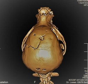
CT Skull: Chihuahua with skull fracture and depressed fractures after a bite injury