Home » Diseases » Emergencies / First Aid for Dogs & Cats – General Information » Emergencies / First Aid A-Z » Foreign Body in the Eye
Foreign Body in the Eye
Foreign bodies in the eyes of dogs and cats represent a common ophthalmological emergency. These are foreign objects that can penetrate various structures of the eye. Our pets’ eyes are complex sensory organs protected from external influences by several natural defense mechanisms such as eyelids, eyelashes, tear fluid, and the blink reflex. The anatomy of the animal eye differs in some aspects from the human eye, which must be considered when examining eye injuries. For example, dogs and cats have a third eyelid (nictitating membrane) that serves as additional protection but can also be affected by foreign bodies itself.
Foreign bodies can lie superficially on the conjunctiva or cornea, be located under the lid or nictitating membrane, or penetrate deeper structures of the eye. Depending on the location, type of foreign body, and duration in the eye, different levels of damage can occur. Animals with protruding eyes, such as brachycephalic breeds (e.g., Pugs, Persian cats), are particularly at risk as their eyes are less protected by the bony eye socket.
Causes
Foreign bodies can easily get into the eye. Smaller foreign bodies are washed out by tears and blinking alone and do not require treatment.
Foreign bodies include in particular:
- Insects
- Grains of sand
- Plant parts
In more detail:
The causes of foreign bodies in the eye are diverse and strongly related to the animal’s living environment and activities. The most common foreign bodies include:
Organic materials such as blades of grass, awns, plant parts, wood splinters, or insects pose a particular danger as they often carry bacteria or fungal spores and can therefore cause infections in addition to mechanical damage. These foreign bodies are particularly common in spring and summer when dogs brush through tall grass or rummage in the undergrowth.
Inorganic materials such as grains of sand, dust, small stones, or metal splinters can also get into the eye, especially during car rides with open windows or in windy conditions. These particles often cause scratches on the cornea, which can lead to severe complications if left untreated.
Chemical substances such as cleaning agents, fertilizers, or other household chemicals can get into the eye if handled improperly and cause severe burns. Particular caution is required here, as the damage is often more serious than with mechanical foreign bodies.
Breed-specific factors also play a role. Dog and cat breeds with protruding eyes or flat faces (brachycephalic breeds) have an increased risk of foreign bodies in the eye, as their eyes are less protected by the skull structure. In addition, some breeds such as Cocker Spaniels or Bloodhounds have drooping eyelids, which can facilitate the entry of foreign bodies.
Symptoms
- Tearing of the eye(s)
- Redness of the conjunctiva
- Blinking
- Keeping the eye closed
If the foreign body penetrates the structures of the eye, the symptoms intensify and lead to
- Pain
- Inflammations
- Swelling of the eyelids
- Clouding of the cornea
The symptoms of foreign bodies in the eye can vary depending on the type, size, and location of the foreign body, but are usually clearly recognizable. Classic signs include increased tearing (epiphora), which is a natural reaction of the body to flush out the foreign body. The conjunctiva often appears red and inflamed (conjunctivitis), which is due to irritation from the foreign body.
Affected animals often show noticeable blinking or keep the eye partially or completely closed (blepharospasm) to reduce pain and avoid further irritation. Typically, they try to rub the affected eye with their paw or rub their head on furniture or the floor, which often worsens the situation and can lead to additional injuries.
If a foreign body remains for a longer period, the symptoms can intensify. Significant swelling of the eyelids (blepharitis) can occur, and the cornea may become cloudy (keratitis). In advanced cases, purulent discharge may form, indicating a secondary bacterial infection. Especially with perforating foreign bodies, inflammation of the inner eye structures (uveitis) can occur, recognizable by a change in pupil size or shape as well as discoloration of the iris.
Some animals may also show increased light sensitivity (photophobia) and actively seek out darker areas. In severe cases, the animal may show signs of general discomfort, such as decreased appetite or reduced activity, especially if pain is pronounced or an infection is present.
First Aid
- Wash your hands with soap and water.
- If the foreign body is larger and you can see it clearly, such as a blade of grass in the area of the conjunctiva, you can try to grasp and remove it or flush it out with warm water. A sterile packed syringe can also be helpful here, which can be used to repeatedly draw up slightly warm, clean tap water and flush it into the eye under gentle pressure.
- Try to keep your animal’s eyelids open while doing this.
- Do not rub the eye, do not apply pressure to the eyeball, and do not use objects to remove a foreign body from the eye.
- Prevent your pet from rubbing its eye with its paws; use a collar if necessary
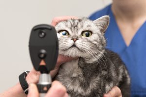
If you notice a firmly lodged foreign object, the eye is significantly reddened, the conjunctiva is already clearly inflamed, or there is even purulent discharge in the corner of the eye, a thorough professional examination of the eyes is necessary (Fig.).
Diagnosis
The diagnosis of foreign bodies in the eye requires a thorough ophthalmological examination. First, the veterinarian conducts a general assessment of the affected eye, looking for signs such as redness, swelling, discharge, and blepharospasm. To enable a detailed examination, a local anesthetic is often administered in the form of eye drops, which alleviates pain and allows the animal to open its eye.
The slit lamp examination is an essential diagnostic tool that provides a magnified, three-dimensional view of the eye. This technique can identify even the smallest foreign bodies or injuries to the cornea. To assess corneal integrity, a fluorescein dye test is often performed. In this test, a fluorescent dye is applied to the eye, which adheres to damaged areas of the cornea and glows green under blue light. This method is particularly valuable for detecting corneal injuries caused by foreign bodies.
If deeper foreign bodies are suspected or in more complex cases, advanced imaging techniques such as ultrasound or even optical coherence tomography (OCT) in specialized facilities may be used. These methods provide a detailed view of the internal eye structures and can help locate foreign bodies that are not visible on the surface.
In some cases, especially when an infection is suspected, swabs may be taken for microbiological examinations. This is important to identify the causative pathogen and initiate targeted antibiotic therapy. When diagnosing, differential diagnoses such as primary conjunctivitis, allergic reactions, or other eye diseases that can cause similar symptoms must also be considered.
Further veterinary measures
Present your pet to a veterinarian.
Superficial foreign bodies can usually be removed under local anesthesia. Foreign bodies that have penetrated into the interior of the eye must be surgically removed.
The treatment of foreign bodies in the eye depends on the type, size, and location of the foreign body, as well as the extent of the damage caused. For superficial foreign bodies on the conjunctiva or cornea, the first step is careful removal. This is done in the veterinary practice under local anesthesia using special instruments such as moist cotton swabs, fine tweezers, or flushing with sterile saline solution. For deeper or larger foreign bodies, sedation or general anesthesia may be necessary to ensure safe removal.
After removal of the foreign body, medical therapy is usually administered to prevent secondary infections and promote healing. Antibiotic eye drops or ointments are prescribed to prevent or treat existing bacterial infections. In cases of severe inflammatory reactions, anti-inflammatory medications such as corticosteroids may also be used, although these must be applied with caution, especially in cases of existing corneal defects.
Pain management is an important aspect of therapy. In addition to local anesthetics for examination and removal of the foreign body, systemic pain medication may be prescribed to provide relief to the animal. In some cases, an Elizabethan collar is recommended to prevent the animal from rubbing its eye and causing further damage.
In cases of perforating injuries or foreign bodies that have penetrated deeper structures of the eye, surgical intervention may be necessary. These procedures are ideally performed by a veterinary ophthalmology specialist. Depending on the severity of the injury, corneal suturing, conjunctival flap surgery, or in severe cases, even removal of the eye (enucleation) may be necessary if it cannot be saved.
Prognosis and aftercare
The prognosis for foreign bodies in the eye depends significantly on several factors: the type and size of the foreign body, the duration of its presence in the eye, the depth of the injury, and the speed of veterinary care. Superficial foreign bodies that are removed early generally have an excellent prognosis. The cornea has a remarkable regenerative capacity, and small superficial injuries often heal completely within a few days without leaving permanent damage.
However, with deeper injuries or prolonged presence of the foreign body, complications can occur. Corneal scars may remain and, depending on their location and extent, can impair vision. Especially in cases of infections with aggressive pathogens or foreign bodies that release chemical substances, healing may be delayed and the risk of permanent damage increases.
Aftercare plays a crucial role in the success of healing. Regular follow-up examinations by the veterinarian are important to monitor the healing process and adjust the therapy if necessary. Correct administration of prescribed medications is essential – eye drops or ointments usually need to be applied several times a day, which can be challenging for pet owners. Precise instructions on the correct technique for administering medication are therefore important.
During the healing phase, the animal should be protected from further injuries. A collar prevents the animal from scratching the eye, and activities that could increase the risk of further eye injuries should be restricted. For brachycephalic breeds or animals with recurring eye problems, long-term preventive measures such as regular eye checks and special care measures may be recommended.
Summary
Foreign bodies in the eye represent a common ophthalmological emergency in dogs and cats that requires prompt action. The range of possible foreign bodies extends from harmless dust particles to dangerous perforating objects such as thorns or metal splinters. Typical symptoms include increased tearing, redness of the conjunctiva, blinking, and partial or complete closure of the eye. If a foreign body remains for an extended period, symptoms can intensify to swelling, corneal opacity, and purulent discharge.
Diagnostic evaluation by a veterinarian is essential and includes a thorough eye examination, often supported by special techniques such as the fluorescein test or slit lamp examination. The therapy depends on the type and location of the foreign body and ranges from simple removal under local anesthesia to complex surgical procedures for deeper foreign bodies. Subsequent medical treatment with antibiotics and anti-inflammatory agents is necessary in most cases.
The prognosis is generally good with early treatment, with the healing duration depending on the severity of the injury. Careful aftercare with regular medication administration and protection from further injuries is crucial for complete recovery. Preventive measures such as avoiding high-risk situations and regular eye checks can help prevent future incidents.
Outlook on current research
Veterinary research in the field of ophthalmology is continuously advancing, including the treatment of foreign bodies in the eye. Innovative imaging techniques such as high-resolution optical coherence tomography (OCT) enable more precise diagnostics and localization of foreign bodies, especially for deeper-lying or very small objects. This technology, originally developed in human medicine, is increasingly being adopted in specialized veterinary facilities and significantly improves diagnostic capabilities.
In the area of therapy, new biomaterials are being researched that can serve as temporary or permanent corneal replacement materials. These could be used in the future for severe injuries caused by foreign bodies to repair the cornea and maintain visual acuity. The development of special contact lenses for animals is also progressing. These can not only serve as protection for the healing cornea but also as carriers for medications that are continuously released over an extended period.
Research on regenerative therapies, including the use of stem cells and growth factors, shows promising results in the treatment of corneal injuries. In the future, these approaches could shorten healing time and reduce the risk of scarring. Furthermore, new antibiotics and anti-inflammatory substances are being developed that act more specifically and cause fewer side effects.
Another important area of research is the development of preventive measures, especially for predisposed breeds. This includes both improved protective devices for animals at increased risk and genetic studies aimed at reducing the breeding of animals with extreme anatomical features that can lead to eye problems.
Frequently asked questions (FAQs)
- How do I know if my pet has a foreign body in its eye?
Look for increased tearing, redness of the eye, frequent blinking, rubbing the eye with the paw, or a partially or fully closed eye. These symptoms indicate pain or discomfort and should be taken seriously. - Can I remove a visible foreign body from my pet’s eye myself?
For superficial, easily visible foreign bodies, you can try to remove them by carefully rinsing with lukewarm, sterile saline solution. However, never use tweezers or other instruments and do not apply pressure to the eyeball. If you are unsure or if the foreign body is not easily removable, seek veterinary help immediately. - How urgent is a veterinary visit if a foreign body in the eye is suspected?
A foreign body in the eye should be considered an emergency. The longer a foreign body remains in the eye, the greater the risk of complications such as infections or permanent damage. Therefore, seek veterinary help within hours if possible. - Which breeds are particularly susceptible to foreign bodies in the eye?
Brachycephalic breeds such as Pugs, Bulldogs, Persian cats, or Pekingese have an increased risk due to their protruding eyes and flat facial shape. Dog breeds with drooping eyelids like Cocker Spaniels or Bloodhounds are also more susceptible to foreign bodies under the lids. - How long does healing take after removing a foreign body from the eye?
The healing duration depends on the severity of the injury. Superficial scratches often heal within 3-5 days, while deeper injuries or infections may require weeks for complete healing. Regular follow-up checks with the veterinarian are important to monitor the healing process. - Can an untreated foreign body in the eye lead to blindness?
Yes, untreated foreign bodies can lead to serious complications such as corneal ulcers, infections, or inflammation of the inner eye structures, which can potentially lead to blindness. - How do I correctly administer eye drops to my pet?
Gently hold your pet’s head, pull the lower eyelid slightly downward, and drip the prescribed amount into the resulting conjunctival sac, not directly onto the cornea. Avoid touching the dropper tip to the eye or fur to prevent contamination. - How can I prevent foreign bodies in my pet’s eye?
Avoid car rides with windows wide open, keep your pet back in strong winds or dusty environments, and regularly check the eyes, especially after walks through tall grass or undergrowth. For predisposed breeds, regular eye care may be beneficial. - Are certain seasons particularly risky for foreign bodies in the eye?
Yes, spring and summer pose an increased risk due to grasses, pollen, and insects. Grass awns are particularly dangerous as they can easily get caught in the eye. In autumn, seeds and dry plant parts can be problematic, while in winter, road salt and grit can cause eye irritation. - Can my pet fully recover after a foreign body injury to the eye?
With early and appropriate treatment, the prognosis for a full recovery is good for most foreign body injuries. Even if scars remain on the cornea, they often do not significantly impair vision unless they are directly in the center of vision.
Literature
- Guideline Professional Association of Ophthalmologists in Germany e.V. and German Ophthalmological Society e.V.,
Guideline No. 8 Injuries to the Eye and its Adnexa, August 2011 with modification regarding chemical burns in February 2024 - Löwe, G. and Löwe, O. (2021). Emergencies in Dogs and Cats – A Veterinary Guide. Kreuztal: Kynos-Verlag.
- Maggs, D.J., Miller, P.E. and Ofri, R. (2017). Slatter’s Fundamentals of Veterinary Ophthalmology. 6th Edition. St. Louis: Elsevier.
- Gelatt, K.N. (2013). Veterinary Ophthalmology. 5th Edition. Ames: Wiley-Blackwell.
- Merck Co. (2020). Merck Veterinary Manual. 11th Edition. Kenilworth: Merck Publishing.
- Ledbetter, E.C. and Gilger, B.C. (2021). Diseases and Surgery of the Canine Cornea and Sclera. In: Gelatt, K.N., Ben-Shlomo, G., Gilger, B.C., Hendrix, D.V.H., Kern, T.J., Plummer, C.E. (Eds.) Veterinary Ophthalmology. 6th Edition. Hoboken: Wiley-Blackwell, pp. 1082-1164.
- Featherstone, H.J. and Heinrich, C.L. (2021). Ophthalmic Examination and Diagnostics. In: Gelatt, K.N., Ben-Shlomo, G., Gilger, B.C., Hendrix, D.V.H., Kern, T.J., Plummer, C.E. (Eds.) Veterinary Ophthalmology. 6th Edition. Hoboken: Wiley-Blackwell, pp. 533-613.
- Gould, D. and McLellan, G.J. (2022). BSAVA Manual of Canine and Feline Ophthalmology. 4th Edition. Gloucester: British Small Animal Veterinary Association.