
Home » Diseases » Emergencies / First Aid for Dogs & Cats – General Information » Emergencies / First Aid A-Z » Joint dislocation (luxation)
Joint dislocation (luxation)
Definition
Dislocation of a joint (luxation)
A luxation is the complete dislocation of a joint with loss of the normal joint position. It causes severe Pain, misalignment, and a marked limitation of function. If left untreated, there is a risk of cartilage, ligament, and nerve damage.
In a “subluxation”, the joint surfaces are still partially in Contact with each other. A temporary luxation is also possible, in which the ends of the bones within a joint shift only temporarily and then return to their physiological position (see –> sprain).
In a luxation, the joint capsule typically tears and surrounding structures such as ligaments, tendons, and in some cases nerves and blood vessels are often damaged. This condition is extremely painful for the affected animal and leads to an immediate functional impairment of the affected joint.
Luxations can occur in both dogs and cats, although the frequency of certain forms of luxation differs between the animal species. In dogs, patellar luxations (kneecap) and hip luxations are particularly common, while jaw and shoulder luxations are more frequently observed in cats. The anatomical features of the respective animal species play a decisive role here. For example, cats have greater joint mobility and elasticity of the ligamentous apparatus, which on the one hand makes their joints more flexible, but on the other hand can also make them more susceptible to certain forms of luxation.
Causes
The causes of luxations in dogs and cats can generally be divided into two categories: traumatic, congenital, and developmental luxations.
Traumatic dislocations are caused by external force, such as those occurring in traffic accidents, falls from great heights (especially in cats), violent fights, or when a limb gets caught. The force applied thereby exceeds the natural resilience of the joint-stabilizing structures. In cats, due to their lifestyle and movement patterns, jaw dislocations after traumas or even during intense Yawning are not uncommon.
Congenital or developmental luxations, on the other hand, are based on genetic factors or growth disorders. Patellar luxation in small dog breeds such as Chihuahuas, Yorkshire Terriers, or Miniature Poodles is a classic example of this. This form of luxation is favored by anatomical malformations such as a too shallow groove for the kneecap or axial deviations of the limbs. In larger dog breeds, severe forms of hip dysplasia can lead to luxations.
Excessive athletic stress can also promote luxations in predisposed animals. In particular, dog sports with rapid changes of direction and jumps such as agility can lead to luxations in anatomically predisposed animals. In cats, repeated jumps from great heights can impair joint stability in the long term.
Breed-specific predispositions also play an important role. In addition to the small dog breeds already mentioned with a tendency to patellar luxation, Labrador Retrievers and German Shepherds, among others, show an increased susceptibility to elbow luxations. Breed-specific predispositions are less pronounced in cats, but certain breeding lines with extreme anatomical features may have an increased risk.
Symptoms
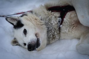
Dislocated joint (luxation)
Typical Symptoms:
- Sudden high-grade lameness, protective posture / guarding posture
- Luxations are very painful
- Abnormal joint position, movement severely restricted or joint can no longer be moved
- Swelling and bruising on and in the joint, tears in the joint capsule vessels
- Partially palpable step formation and cavity in the joint area
- Often after jump/trauma
Alarm signs:
- Non-weight bearing + significant malalignment
- Severe pain, swelling, possible vascular/nerve damage
- Open wound on the joint or accident with multiple injuries
Escalation/course:
- Hours: Swelling/pain make repositioning difficult
- Days: Muscle shortening/instability → prognosis worse
- Repeated dislocation → chronic instability
The clinical signs of a luxation are usually acute and pronounced. Affected animals typically show a sudden onset of high-grade lameness to complete relief of the affected limb. However, the symptomatology can vary depending on the affected joint and extent of the injury.
The following characteristic symptoms can be observed in dogs and cats with a luxation: The affected joint appears fixed in an unnatural position and can no longer be moved physiologically. Tearing of blood vessels in the joint capsule often results in significant swelling and bruising in the joint area. On palpation, the veterinarian can often detect a step formation or an abnormal cavity in the joint area, as the joint surfaces are no longer correctly aligned.
The expression of pain varies between animal species. While dogs often indicate pain by whining, howling, or aggressive behavior when the affected area is touched, cats often show more subtle signs such as withdrawal, decreased activity, or hissing when approaching the painful region.
In chronic or recurrent luxations, such as those that can occur with patellar luxation, the symptoms are less dramatic. Here, one often observes an intermittent pulling up of the affected limb for a few steps, followed by normal weight-bearing when the kneecap spontaneously slides back into its position. Cats with chronic jaw luxation typically show difficulty eating and drool more.
Especially in traumatic luxations, accompanying symptoms such as shock, increased heart rate, shallow breathing, and pale mucous membranes may occur, requiring immediate veterinary emergency care.
Special considerations for cats
Cats often express pain ‘silently’ (withdrawal, unusual quietness). Therefore, a lower threshold for a ‘Red’ alert applies. Always consider it ‘Red’ if the breathing rate is increased, mouth breathing, and/or low body temperature (hypothermia) are present.
After a fall or significant trauma, chest and internal injuries are often present despite apparent stability.
First Aid
- The joint should no longer be moved.
- Do not attempt to put the joint back into the correct position. It could cause further damage to the cartilaginous joint surfaces as well as to nerves, blood vessels, and ligaments in the region.
- You can reduce the swelling of the joint by cooling it with a wet bandage until you arrive at the veterinarian.
- Correcting a luxation is a matter for a veterinarian. Do not attempt to manipulate the joint yourself!
When should you see a vet sooner?
Urgency increases from orange to → red in cases of very severe Pain (unmanageability), clear deformity after an accident (suspected fracture-dislocation), an open wound over the joint, or neurological deficits (paw dragging/no reaction).
Diagnosis
The diagnosis of a luxation is made by a systematic clinical examination in combination with imaging procedures. First, the veterinarian performs a thorough medical history to obtain information about the accident or pre-existing problems. The observation of the gait – if the animal can still walk – gives initial indications of the affected limb and the extent of the functional impairment.
During the orthopedic examination, the veterinarian carefully palpates the affected joint and checks the joint mobility. Due to the severe pain, this examination is often performed under sedation or anesthesia to avoid additional stress and pain for the animal. The veterinarian pays attention to abnormal joint positions, instabilities, and pain.
Imaging techniques are essential for a definitive diagnosis. X-rays in at least two planes are the gold standard and allow the assessment of the joint position as well as the exclusion of accompanying fractures. In more complex cases or for a more detailed representation of soft tissue structures, further imaging procedures such as computed tomography (CT) or magnetic resonance imaging (MRI) can be used. These procedures are particularly valuable for assessing the extent of damage to ligaments, tendons, and cartilage surfaces.
In chronic or recurrent luxations, such as patellar luxation, a classification according to severity is also carried out. In patellar luxation, a distinction is made between four grades:
- Grade 1: The kneecap can be manually luxated but springs back on its own
- Grade 2: The kneecap luxates occasionally but can be manually repositioned
- Grade 3: The kneecap is permanently luxated but can be manually repositioned
- Grade 4: The kneecap is permanently luxated and can no longer be repositioned
In some cases, a diagnostic arthroscopy may also be useful to directly visualize the extent of joint damage and initiate therapeutic measures at the same time.
Further veterinary measures
A careful clinical examination of traumatic luxations provides indications as to whether ligaments are injured and whether nerves or blood vessels are additionally damaged.
This is usually followed by further examinations of the joint under general anesthesia and several X-rays.
If no stability can be achieved after repositioning the joint parts in the correct anatomical position, surgery is necessary.
The unstable position of the kneecap, which is usually genetically determined in small breeds, occupies a certain special position. The cause is altered anatomical conditions, which can be surgically stabilized very successfully with different methods depending on the initial situation.
Supplements
The treatment of luxations depends on the type of joint affected, the cause of the luxation, the severity, and the duration since it occurred. Basically, a distinction is made between conservative and surgical therapy approaches.
For fresh, uncomplicated dislocations, a closed reduction under anesthesia can be attempted. This involves repositioning the dislocated joint to its physiological position through targeted manipulation. Afterwards, stabilization is performed with a bandage, a splint, or a cast for several weeks to allow the injured ligamentous apparatus time to heal. This method is particularly successful for shoulder joint dislocations in cats and some forms of hip joint dislocation.
In more complex luxations, repeated occurrences, or if there are accompanying injuries such as ligament ruptures or cartilage damage, surgical intervention is necessary. The surgical techniques vary depending on the joint affected:
In patellar luxation, depending on the severity, the surgical measures include deepening the groove for the kneecap (trochleoplasty), displacing the tibial tuberosity (to correct the direction of pull of the quadriceps muscle), or plication of the joint capsule. In hip luxations, various techniques such as capsule plication, insertion of artificial ligaments, or, in severe cases, artificial hip replacement can be used.
Modern minimally invasive techniques are becoming increasingly important. Arthroscopy-assisted procedures enable less tissue trauma and faster rehabilitation. In complex cases, individualized implants can be manufactured using 3-D printing technology to ensure optimal anatomical fit.
Drug therapy accompanies both conservative and surgical treatments and includes analgesics for pain relief, anti-inflammatory drugs to reduce inflammation, and in some cases muscle relaxants. In cats, special care must be taken when selecting medications, as they metabolize certain active ingredients such as non-steroidal anti-inflammatory drugs less well.
An essential component of the therapy is physical rehabilitation, which ideally begins in the early postoperative phase. Controlled movement exercises, hydrotherapy on the underwater treadmill, laser therapy, and targeted massage techniques support the restoration of joint function and the building of the stabilizing muscles.
Prognosis and aftercare
The prognosis after a luxation depends on various factors, including the affected joint, the cause of the luxation, the time between onset and treatment, and the presence of concomitant injuries. In general, the earlier the treatment, the better the prognosis.
In fresh, traumatic luxations without significant concomitant injuries, the prognosis after correct repositioning and adequate aftercare is usually good to very good. For example, shoulder luxations in cats have a success rate of over 80% after successful closed repositioning. In patellar luxations of grades 1 and 2, a complete restoration of joint function can be expected in about 90% of cases after surgical correction.
Complicated dislocations with extensive soft tissue damage, accompanying fractures, or existing degenerative changes have a more guarded prognosis. There is an increased risk of developing post-traumatic osteoarthritis or chronic instability.
Aftercare plays a crucial role in the success of the treatment and includes several components:
Movement restriction in the early phase after reduction or surgery is essential to enable the healing of the injured structures. Depending on the affected joint and the type of treatment, this phase can last between two and eight weeks. During this time, dogs should only be walked on a short leash, while cats should ideally be kept in a confined space.
Structured rehabilitation begins with passive range of motion exercises and slowly progresses to active exercises and controlled increases in weight-bearing. An individually tailored rehabilitation program can shorten the healing time and improve the functional outcome. Modern rehabilitation concepts include hydrotherapy, laser therapy, therapeutic ultrasound, and targeted massage techniques.
Regular veterinary check-ups with clinical examinations and, if necessary, follow-up X-rays are important to monitor the healing process and detect complications early. Particular attention is paid to joint stability, range of motion, and signs of pain or inflammatory conditions.
Weight management is an often underestimated aspect of aftercare. Overweight puts additional stress on the joints and can delay the healing process or increase the risk of renewed dislocations. An adapted diet with optimal calorie intake and, if necessary, the use of joint-supporting nutritional supplements such as glucosamine, chondroitin, and omega-3 fatty acids can support the healing process.
Summary
Dislocations in dogs and cats are a common orthopedic condition that can occur both traumatically and developmentally. The complete displacement of the joint surfaces leads to severe pain and functional limitations. While patella and hip dislocations are common in dogs, jaw and shoulder dislocations are more frequently observed in cats.
Diagnostics include a thorough clinical examination and imaging techniques, with X-rays being the gold standard. The therapy depends on the type and severity of the dislocation and can range from closed reduction with subsequent immobilization to complex surgical procedures. Modern surgical techniques and minimally invasive procedures have significantly improved treatment outcomes in recent years.
The prognosis is usually good with early and adequate treatment, with consistent aftercare and structured rehabilitation having a decisive influence on the functional outcome. In the long term, severe dislocations carry the risk of post-traumatic osteoarthritis, which requires continuous monitoring and, if necessary, symptomatic therapy.
Preventive measures such as weight control, adapted physical activity, and responsible breeding selection for hereditary forms of dislocation can reduce the risk of dislocations. Close cooperation between pet owners, veterinarians, and physical therapists is the key to an optimal treatment outcome and a good quality of life for the affected animals.
Outlook on current research
Research in the field of dislocation treatment in dogs and cats is constantly evolving. Current studies focus on various innovative approaches to improve both diagnostics and therapy.
In the field of imaging diagnostics, new high-resolution MRI protocols enable a more detailed representation of soft tissue structures such as ligaments, tendons, and cartilage. This improved imaging helps to detect subtle damage and optimize therapy planning. Dynamic examination techniques also allow the assessment of joint function under load, which is particularly important in intermittent dislocations.
In the surgical field, biomechanically optimized implants are increasingly being developed to enable more stable fixation with less tissue irritation. The use of biocompatible materials with bone-like mechanical properties reduces the risk of implant loosening and failure. Personalized implants, manufactured using 3-D printing technology based on CT data of the individual patient, enable an optimal anatomical fit and improved functionality.
Regenerative therapy approaches are becoming increasingly important. The use of stem cells, platelet-rich plasma (PRP), and growth factors aims to promote the healing of damaged cartilage and ligaments. Initial clinical studies show promising results, especially in the treatment of accompanying cartilage damage after dislocations.
Genetic research is making progress in identifying genes associated with hereditary forms of dislocation, primarily patella dislocation. In the future, this could lead to genetic tests that enable more targeted breeding selection and reduce the prevalence of these diseases.
In the field of rehabilitation, computer-assisted gait analysis and pressure measurement plates are increasingly used to objectively document the rehabilitation process and individually adapt the therapy. Virtual reality and robot-assisted therapy systems, which are already established in human medicine, are being adapted for veterinary use.
The development of minimally invasive techniques is progressing. Arthroscopically assisted reconstructions of ligaments and capsule structures enable less tissue trauma and faster rehabilitation. These techniques are continuously refined and extended to other joints and indications.
Despite these promising developments, there is still a need for research, primarily with regard to the long-term results of new therapy approaches and the optimization of prevention strategies. Interdisciplinary collaboration between veterinarians, biomechanics, materials scientists, and geneticists will be crucial to further improve the treatment of dislocations in dogs and cats.
Frequently asked questions (FAQs)
-
What is meant by a luxation in dogs?
A luxation (joint dislocation) is an injury in which the bones of a joint are completely or partially (subluxation) displaced from their normal position. The joint capsule and surrounding structures such as ligaments or tendons can be damaged. Commonly affected joints in dogs are the shoulder, hip, knee, and elbow joints. A luxation differs from a sprain or strain in that the joint surfaces no longer meet normally. -
What are the causes and risk factors for a luxation in dogs?
- Traumatic events: Falls, collisions with vehicles, violent brawls, or sports injuries can trigger a luxation.
- Congenital malformations: Some breeds have congenital anatomical features that lead to unstable joints (e.g. patellar luxation in smaller breeds).
- Overweight: Excessive body weight puts additional stress on the joints and can increase the risk of injury.
- Degenerative diseases: Wear and tear (e.g. osteoarthritis) or ligament and tendon weakness can impair stability in the joint and promote luxation.
-
What symptoms indicate a luxation?
- Sudden lameness or unwillingness to put weight on the affected leg
- Severe pain with movements in the corresponding joint
- Abnormal position of the limb (e.g. twisted or crooked leg)
- Swelling or hematomas around the joint
- Appeasement behavior (licking, guarding posture, whining) or aggressiveness when touched
- A typical sign can be an audible cracking sound during the accident or the absence of normal joint mobility.
-
How is a luxation diagnosed and treated?
- Clinical examination: The veterinarian checks the gait, carefully palpates the affected joint, and looks for instability.
- Imaging procedures: X-rays are the gold standard to confirm a luxation and rule out possible bone fractures. In addition, an ultrasound or CT scan may be useful if soft tissues (e.g. ligaments, menisci) need to be examined more closely.
- Treatment methods:
- Reposition: The joint is carefully brought back into the anatomically correct position (often under sedation or anesthesia).
- Splinting/stabilization: Depending on the severity of the injury, the joint is immobilized or stabilized with the help of ligaments, sutures, or implants.
- Surgery: In severe luxations, complicated ligament injuries, or recurrent luxations, a surgical procedure may be necessary. Here, the joint is reconstructed and stabilized, e.g. by artificial ligaments or by displacing bone attachments.
- Physiotherapy: Physiotherapy is often recommended to support healing and restore joint function.
-
How can you prevent a luxation in dogs?
- Weight control: A healthy body weight reduces the stress on the joints.
- Sufficient exercise: Moderate, regular training strengthens the muscles, ligaments, and tendons.
- Note breed-specific risks: In breeds with a tendency to patellar luxation or hip dysplasia, targeted muscle building training and a regular veterinarian check are useful.
- Avoid strong load peaks: Too rapid turns, jumps from great heights, or excessive athletic stress can increase the risk of injury.
- Early detection: At the first signs of lameness or pain, a veterinarian should be consulted to intervene early before severe joint damage or luxations occur.
Conclusion: Luxations are painful and potentially consequential joint injuries in dogs that require rapid action. Professional diagnosis and therapy by the veterinarian are crucial to stabilize the affected joint and avoid long-term complications.
Literature
- Löwe, G. and Löwe, O. (2021). Emergencies in dogs and cats – A veterinary Guide. Kynos-Verlag. 208 pp.
- Schulz KS, Hayashi K, Fossum TW. Diseases of the joints. In: Fossum TW, ed. Small Animal Surgery. 5th ed. Philadelphia: Elsevier; 2019:1134-1279.
- DeCamp CE, Johnston SA, Déjardin LM, Schaefer SL. Handbook of Small Animal Orthopedics and Fracture Repair. 5th ed. St. Louis: Elsevier; 2020.
- Witte PG, Scott HW. Conditions of the feline patellar ligament in cats with and without medial patellar luxation. Veterinary and Comparative Orthopaedics and Traumatology. 2020;33(3):177-181.