
Laceration
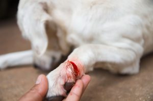 Dog with a deep laceration
Definition
Laceration
A laceration is a sharply defined wound caused by glass, metal, or sharp edges. It can be superficial or affect tendons, vessels, and nerves. Severe Bleeding, gaping edges, or deep structures make it relevant to emergencies.
Severe lacerations in pets are a serious medical emergency that requires immediate action. In contrast to superficial cuts, severe lacerations are characterized by a deeper penetration of the skin and underlying tissues. These injuries are classified into four grades according to their depth and the extent of the affected structures: Grade I affects only the epidermis, Grade II extends into the dermis, Grade III affects the subcutaneous tissue, and Grade IV affects deeper structures such as muscles, tendons, nerves, or blood vessels.
Severe lacerations in anatomical risk zones such as the neck, chest, abdomen, or on large joints are particularly dangerous. In these areas, vital structures can be affected, which can lead to life-threatening complications such as severe Blood loss, infections, or functional impairments. Knowledge of these basics is crucial for correct first aid and assessment of the urgency of veterinary treatment.
Causes
Lacerations in the paw area are not uncommon. Bleeding may occur. This usually stops on its own after a short time.
Small lacerations with smooth edges usually heal quickly.
Serious lacerations occur, for example, when jumping into broken glass or onto an upright bottle bottom. This can lead to deep lacerations involving blood vessels, tendons, ligaments, and joints.
Supplements
The causes of severe lacerations in pets are varied. In the home environment, shards of glass, sharp-edged metal objects, and kitchen utensils are common sources of danger. Cats, in particular, can suffer severe lacerations when passing through windows or climbing over fences. In dogs, these injuries often occur when running over broken glass, scrap metal, or sharp-edged stones.
In rural areas, agricultural equipment, barbed wire, and metal fences are common causes. Hunting dogs often suffer injuries from thorns, sharp undergrowth, or game fences. Traffic accidents can also lead to complex lacerations in which glass or metal splinters penetrate deep into the tissue.
A particular risk is posed by modern household appliances with sharp edges or blades, such as lawnmowers, fans, or kitchen appliances. These can lead to particularly serious injuries in unsupervised animals, as the cuts are often deep and made with considerable force.
Symptoms
Laceration
Typical Symptoms:
- Visible wound, smooth wound edges
- Linear transection of the skin and possibly underlying layers
- Depending on depth: gaping, fatty tissue/tendons visible
- More or less severe Bleeding, sometimes pulsating
- Lameness due to Pain if extremities are affected
Alarm signs:
- Severe Bleeding, pulsating/spurting
- Visible tendon/nerve/bone, loss of function
- Wound on the neck/thorax / chest/abdomen or near a joint
- Signs of shock
Escalation/course:
- Minutes: Blood loss can become critical
- Hours: Contamination/risk of infection increases
- Days: Abscess, necrosis, suture dehiscence possible
The symptoms of severe lacerations vary depending on the location and severity. The most obvious sign is a linear transection of the skin with smooth or frayed wound edges. In deep cuts, the wound edges gape open, with deeper structures such as fatty tissue, muscles, or even tendons becoming visible.
Severe Bleeding is a characteristic feature of severe lacerations, especially when arteries are affected. In this case, the Bleeding is bright red and pulsating. In the case of venous injuries, the blood is dark red and flows more evenly. In addition to the Bleeding, affected animals often show Pain, which can be expressed through Lameness, Protective posture / guarding posture, Licking the wound, or vocalizations.
If nerves are injured, neurological deficits such as loss of sensation or restricted movement may occur. If tendons are severed, the animal can no longer put normal weight on or move the corresponding limb. In advanced injuries or beginning infections, Swelling, redness, and increased body temperature may occur. In severe cases, shock symptoms such as pale mucous membranes, increased heart rate, and weakness may occur.
Special considerations for cats
Lick protection is important. Cat wounds can be deeper than they appear.
First Aid
- Allow minor lacerations to bleed slightly for self-cleaning
- After washing your hands, apply pressure to the wound with a clean cloth until the Bleeding stops
- Clean contaminated wounds with fresh, preferably running tap water.
- Wash the surrounding area with soap
- Cover small wounds with an antibiotic ointment
- Protect the wound from dirt with a light bandage that must be changed daily
- In the case of deep wounds or wounds where the Bleeding does not stop despite applying pressure, arteries may be injured and a bandage should always be applied
- If blood still comes out, put another bandage over it
- In the case of deeper lacerations just above the paws, tendons are also often injured. Do not apply a tourniquet over tendons.
- After applying a bandage, you should see a veterinarian, as tendons must be treated surgically and never heal on their own.
- For deep wounds, veterinary care reduces the risk of infections, including tetanus infection.
- If the surrounding area becomes inflamed in the case of apparently small wounds or if you feel unsure, you should also present your animal to a veterinarian.
When should you see a vet sooner?
Urgency increased from orange to → red in cases of heavy Bleeding, deep Wounds, or visible structures (muscles/tendons/bones).
Diagnosis
The diagnosis of severe lacerations begins with a thorough clinical examination. The veterinarian first assesses the extent of the injury, the depth of the cut, and the structures that may be affected. The examination includes the assessment of the wound edges, the examination for foreign bodies, and the evaluation of the surrounding tissues.
Imaging procedures are often necessary for deep lacerations. X-rays can identify foreign bodies such as glass or metal splinters and rule out bone injuries. In the case of complex injuries in the area of joints or if tendon injuries are suspected, an ultrasound examination can provide valuable information. In particularly severe cases, CT or MRI examinations may be necessary to assess the full extent of the tissue damage.
Laboratory tests supplement the diagnosis, especially in the case of severe Blood loss or suspected infections. A blood count provides information about the severity of the Blood loss and can provide indications of inflammatory processes. In the case of infected Wounds, a bacteriological examination with an antibiogram is useful to determine the appropriate antibiotic therapy.
Further veterinary measures
Deeper, very long or gaping lacerations are stapled or sutured under a short general anesthesia (Fig.).
If tendons, ligaments, or joints are injured, they must be surgically repaired in the majority of cases.
In the case of such injuries, a stabilizing bandage is necessary after the operation.
The healing process of tendons takes several weeks.
If blood vessels and nerves are injured at the same time, permanent functional impairments can occur. Reruptures (re-tearing of the sutured tendon) are also possible.
The healing of tendons and ligaments can be supported by methods of regenerative therapy.
Supplements
The therapy of severe lacerations requires a multi-stage approach. After stabilization of the patient and control of life-threatening Bleeding, thorough wound cleaning is performed under sterile conditions. Foreign bodies are removed and dead tissue is surgically removed (debridement).
In the case of deep lacerations, surgical treatment under general anesthesia is usually necessary. Severed blood vessels are ligated or sutured, injured nerves are adapted, and severed tendons or muscles are reconstructed using special suture techniques. The wound is closed in layers, with deeper structures being closed with absorbable suture material and the skin with non-absorbable material or staples.
In the case of heavily contaminated wounds or injuries with significant tissue loss, the insertion of drains may be necessary. In particularly severe cases, modern wound care techniques such as negative pressure wound therapy (vacuum therapy) are used. Drug therapy includes antibiotics for infection prophylaxis, analgesics for Pain relief, and, if necessary, tetanus prophylaxis.
Prognosis and aftercare
The prognosis for severe lacerations depends largely on the location, extent of the injury, and speed of treatment. Superficial, clean cuts have an excellent prognosis with correct care. In the case of deep injuries involving tendons, nerves, or joints, the prognosis should be more cautious, as permanent functional impairments are possible.
Aftercare plays a crucial role in the healing process. Regular bandage changes under sterile conditions are necessary to monitor the healing process and identify complications early on. Wound control includes the assessment of the wound edges, the examination for signs of infections, and the evaluation of the granulation tissue.
Controlled rehabilitation is important for injuries to tendons or joints. This can include passive range of motion exercises, controlled load increase, and, if necessary, physiotherapeutic measures. The duration of aftercare varies depending on the severity of the injury and can take several months in complex cases.
Summary
Severe lacerations in pets are a serious medical challenge that requires immediate action. The classification according to severity and the identification of affected anatomical structures are crucial for therapy planning. The most common causes are accidents with shards of glass, metal objects, or sharp-edged household appliances.
The symptomatology ranges from visible tissue transections and Bleeding to functional impairments and shock symptoms. The diagnosis is based on clinical examination, imaging procedures, and, if necessary, laboratory tests. The therapy includes surgical wound care, the reconstruction of injured structures, and accompanying drug treatment.
The prognosis depends on the extent of the injury and the quality of care. Careful aftercare with regular check-ups and, if necessary, rehabilitative measures is essential for an optimal result. Through early detection and professional treatment, good functional results can often be achieved even in severe lacerations.
Outlook on current research
Research in the field of treatment of severe lacerations in pets is constantly evolving. Innovative approaches such as the use of autologous growth factors and platelet-rich plasma (PRP) show promising results in promoting wound healing and regeneration of tendon and muscle tissue. These biological therapies use the body’s own healing mechanisms and can shorten the healing time and improve functional results.
Another focus of research is on the development of advanced wound dressings and biomaterials. Modern wound dressings with antimicrobial properties, controlled drug release, or integrated sensors for monitoring the wound environment are under development. Bioactive materials that promote tissue regeneration and serve as a scaffold for cell colonization represent a promising approach for the treatment of complex Wounds.
Stem cell therapy is also gaining increasing importance in veterinary medicine. Mesenchymal stem cells can promote wound healing, modulate inflammatory reactions, and support tissue regeneration. First clinical studies show positive results in the application for complex Wounds and tendon injuries.
Frequently asked questions (FAQs)
- How do I know if a laceration in my pet is considered severe?
A severe laceration is characterized by deep tissue transection, severe Bleeding, gaping wound edges, or visible deeper structures such as fatty tissue, muscles, or tendons. Functional impairments of the affected body region also indicate a severe injury. - What should I do if my pet suffers a severe laceration?
Stay calm, stop severe Bleeding by applying pressure with clean cloths, and transport your animal to the veterinarian immediately. Avoid home remedies or your own treatment attempts for deep Wounds. - How long does it take for a severe laceration to heal?
The healing time varies depending on the severity, location, and affected structures. Superficial cuts can heal in 7–14 days, while deep injuries involving tendons or nerves can take several months to heal completely. - What complications can occur with severe lacerations?
Possible complications include infections, impaired wound healing, suture dehiscence, seromas, hematomas, permanent functional impairments, Scar tissue formation, and reruptures in the case of tendon injuries. - Are antibiotics always necessary for severe lacerations?
Antibiotics are usually prescribed for deep or heavily contaminated Wounds to prevent infections. The veterinarian makes the decision based on the individual case. - How can I prevent my pet from Licking or scratching the wound?
A neck collar (Elizabethan Collar), special bodysuits, or bandages can prevent the animal from manipulating the wound. The appropriate method depends on the location of the injury. - Can a severe laceration lead to permanent damage?
Yes, especially in the case of injuries to nerves, tendons, or joints, permanent functional impairments can remain despite optimal treatment. - How do I recognize signs of infection in a healing laceration?
Increasing redness, Swelling, warmth, increased or foul-smelling discharge, Pain, and systemic symptoms such as Fever or loss of appetite may indicate an infection.
Literature
- Löwe, G. and Löwe, O. (2021). Emergencies in dogs and cats – A veterinary Guide. Kreuztal: Kynos-Verlag. 208 pp.
- Pavletic MM, Trout NJ. Bullet, bite, and burn wounds in dogs and cats. Veterinary Clinics of North America: Small Animal Practice. 2019;49(5):895-912.
- Bohling MW, Henderson RA. Differences in cutaneous wound healing between dogs and cats. Veterinary Clinics of North America: Small Animal Practice. 2021;51(4):759-778.
- Hosgood G. Open wounds. In: Williams J, Moores A, editors. BSAVA Manual of Canine and Feline Wound Management and Reconstruction. 2nd ed. Gloucester: British Small Animal Veterinary Association; 2022. p. 37-58.
- Tambella AM, Attili AR, Dupré G, et al. Management of wound healing and complications in surgical wounds in the dog and cat. Animals. 2020;10(9):1505.