Bruising
A bruise, medically referred to as a hematoma, occurs when blood accumulates outside the blood vessels in the tissue. During this process, blood vessels rupture, causing blood to leak into the surrounding tissue and collect there. Hematomas can occur both superficially under the skin (subcutaneous) and in deeper layers of tissue such as muscles, organs, or body cavities. In dogs and cats, we distinguish between different types of hematomas, which differ in their location and formation. Particularly common are subcutaneous hematomas, which form directly under the skin and become visible as bluish-purple discolorations, as well as aural hematomas, which specifically occur on the ear flap. The formation of a hematoma is part of the body’s own response to tissue injury and represents the first step in the healing process. The leaked blood initially clots and is then reabsorbed by the body over time, with characteristic color changes occurring from dark red to blue-purple to yellowish-green.
Causes
Bruises in pets occur through various mechanisms that all lead to damage of blood vessels. The most common cause is blunt trauma such as impacts, falls, or collisions, where the skin remains intact but underlying vessels rupture. In dogs, hematomas often occur after fights with other dogs, traffic accidents, or by forcefully bumping into furniture. Cats often suffer bruises from falls from great heights or during territorial fights. A special form is the aural hematoma, which occurs due to vigorous head shaking during ear infections or foreign bodies in the ear canal, where blood vessels between the ear cartilage and skin rupture. Surgical procedures can also lead to hematomas, especially if hemostasis during the operation was insufficient or if there is increased activity postoperatively. In older animals or animals with certain underlying conditions, hematomas can also occur spontaneously or after minimal trauma. Underlying causes can include coagulation disorders such as von Willebrand’s disease, thrombocytopenias, liver diseases, or the intake of anticoagulant medications. Tumor diseases, especially hemangiosarcomas, can also lead to spontaneous bleeding and hematomas.
Symptoms
The clinical signs of a hematoma vary depending on location, size, and age of the injury. Superficial subcutaneous hematomas manifest as visible, initially reddish-purple, later greenish-yellow discolorations of the skin. They are often associated with swelling, which can vary in intensity depending on the extent of bleeding. On palpation, fresh hematomas feel firm-elastic and are typically painful. Animals often show lameness or a protective posture if limbs are affected. With ear hematomas, a characteristic fluctuating, doughy swelling of the ear flap occurs, which can lead to deformation. Affected animals often shake their heads or scratch their ears more frequently. Deeper hematomas in muscles or organs may not be visible externally and manifest through non-specific symptoms such as reluctance to move, pain on touch, or functional limitations of the affected organ. With large or massive bruises, significant blood loss can occur, manifesting in pale mucous membranes, increased heart rate, and weakness. As healing progresses, the appearance of the hematoma changes: The swelling decreases, the color changes from dark red through blue-purple to green-yellow, and the pain subsides.
First Aid
- For smaller bruises, treatment is often not necessary.
- Immediate cooling (ice pack) for 10 to 20 minutes, possibly multiple times within 24 hours, and rest, at least for the next few hours, prevents or slows further blood loss from the injured blood vessel.
- For a developing severe bruise, you can also apply a pressure bandage for a short time (maximum 20 minutes) while simultaneously cooling.
- After that, however, you should remove the bandage, as often the experience is lacking in how strong the applied pressure should be. The blood flow and thus nutrition of the tissue must not be prevented!
- It’s better to leave head bandages for an aural hematoma to your veterinarian.
- Do not open an aural hematoma on your own.
Diagnosis
The diagnosis of a hematoma is primarily made through clinical examination. The veterinarian looks for typical signs such as swelling, skin discoloration, and tenderness upon palpation. The medical history plays an important role in identifying possible causes such as trauma or underlying diseases. For superficial hematomas, the diagnosis is usually straightforward. For deeper hematomas or to determine the extent, imaging techniques may be necessary. Ultrasound examinations allow for the visualization of fluid accumulations in the tissue and help to assess the size of the hematoma as well as possible involvement of deeper structures. X-rays may be indicated if bone fractures or organ injuries are suspected. In more complex cases, computed tomography (CT) or magnetic resonance imaging (MRI) are used. For recurrent or spontaneously occurring hematomas, further diagnostic tests should be performed to identify possible underlying diseases. These include blood tests to assess coagulation function (platelet count, coagulation times), liver diagnostics, and if necessary, specific tests for coagulation disorders such as von Willebrand factor. Fine needle aspiration or biopsy of the hematoma may help differentiate from other swellings such as tumors or abscesses in unclear cases.
Further veterinary measures
The treatment of bruises depends on size, location, and severity. For smaller, superficial hematomas, conservative therapy is often sufficient. In the acute phase (first 24-48 hours), cold therapy is the focus: Applying cold compresses for 15-20 minutes several times a day promotes vasoconstriction and minimizes further blood loss. After 48 hours, heat applications can be used to accelerate the resorption of the hematoma. For pain relief, non-steroidal anti-inflammatory drugs such as carprofen or meloxicam are used, while monitoring kidney function. For larger or problematic hematomas, puncture and drainage may be necessary to remove accumulated blood and reduce pressure on surrounding tissue. Aural hematomas usually require surgical intervention, as they are rarely resorbed spontaneously. Here, the accumulated blood is removed and sutures or drains prevent re-accumulation. At the same time, the underlying cause (e.g., ear infection) should be treated. For hematomas due to coagulation disorders, the underlying disease must be treated, for example, by vitamin K substitution in cases of poisoning by anticoagulants or specific treatments for thrombocytopenias. In severe cases with significant blood loss, blood transfusions may be necessary. Immobilization of the affected body part is an important part of the therapy to prevent further bleeding.
Prognosis and aftercare
The prognosis for bruises is generally favorable in most cases. Smaller, superficial hematomas usually heal completely within 2-3 weeks without leaving permanent damage. However, the healing time can vary depending on the size, location, and underlying conditions of the animal. Larger hematomas or those in sensitive areas may take longer to heal. Aural hematomas tend to recur without adequate surgical treatment or can lead to permanent deformities of the ear flap. Aftercare includes regular monitoring of the healing process, paying attention to signs of infection such as increasing redness, warmth, or purulent discharge. For surgically treated hematomas, dressing changes and suture removal should be carried out according to veterinary instructions. The animal’s activity should be restricted during the healing phase to avoid new trauma. Particularly important is the identification and treatment of underlying diseases in recurring or spontaneously occurring hematomas. A protective collar may be necessary to prevent the animal from licking or biting the affected area. For aural hematomas, aftercare is especially important to prevent recurrence. This includes consistent treatment of ear infections and regular ear hygiene.
Summary
Bruises (hematomas) are common injury consequences in pets, occurring when blood leaks into the tissue following vessel damage. They can occur superficially under the skin or in deeper tissue layers and typically manifest as painful swellings with characteristic discoloration. The most common causes are blunt trauma, while for aural hematomas, specifically vigorous head shaking due to ear infections is causal. Diagnosis is primarily clinical but can be supplemented by imaging procedures and laboratory tests, especially when underlying conditions are suspected. Therapeutically, conservative treatment with cooling, pain medication, and immobilization is the focus for smaller hematomas, while larger or problematic hematomas may require drainage or surgical intervention. The prognosis is generally good, with healing time depending on various factors. Careful aftercare and treatment of underlying diseases are important to avoid complications and recurrence. Special attention should be paid to recurring or spontaneously occurring hematomas, as they may indicate coagulation disorders or other systemic diseases.
Outlook on current research
Current veterinary research on hematomas focuses on several promising areas. Innovative treatment approaches such as the use of Platelet-Rich Plasma (PRP) show positive effects on healing speed and quality of tissue regeneration in initial studies. This therapy utilizes the body’s own growth factors from concentrated platelets to accelerate healing processes. In the field of aural hematomas, new minimally invasive surgical techniques are being developed to enable faster healing with less scarring. Laser-assisted procedures and special drainage systems are being evaluated to reduce recurrence rates and achieve better cosmetic results. The molecular mechanisms of hematoma resorption are being researched more intensively to develop pharmacological interventions that can accelerate natural breakdown. Here, anti-inflammatory substances and enzymes that specifically promote the breakdown of blood components are in focus. Advances in diagnostic imaging, particularly high-resolution ultrasound techniques and functional MRI technologies, allow for more precise assessment of hematomas and their effects on surrounding tissue. This contributes to individualized treatment strategies. Genetic studies on coagulation disorders in various dog and cat breeds help identify at-risk populations and develop preventive measures. These research approaches promise improved diagnostic and therapeutic options for hematomas in pets in the coming years.
Frequently asked questions (FAQs)
- How Long Does It Take for a Hematoma to Heal in My Pet?
Smaller hematomas typically heal within 2-3 weeks. For larger bruises or those in unfavorable locations, healing can take 4-6 weeks or longer. - Can I Treat a Hematoma in My Pet Myself?
Small, superficial hematomas can often be managed at home by cooling in the first 24-48 hours followed by rest. For larger hematomas, severe pain, or hematomas in sensitive areas, a veterinarian should always be consulted. - Why Do Some Dogs Develop Recurring Othematomas?
Recurring othematomas often result from chronic ear infections, allergies, or foreign bodies that lead to increased head shaking. A thorough diagnosis and treatment of the underlying cause are crucial. - When Is a Hematoma in My Pet an Emergency?
A hematoma becomes an emergency if it rapidly increases in size, is associated with severe pain, impairs breathing or other vital functions, or shows signs of significant blood loss (pale mucous membranes, weakness). - Can Hematomas in Pets Burst on Their Own?
Yes, large superficial hematomas can occasionally rupture spontaneously, especially if they are under tension or if the animal licks/scratches at them. This carries infection risks and should be treated by a veterinarian. - Are There Breeds More Prone to Hematomas?
Yes, breeds with known coagulation disorders such as Dobermans (von Willebrand’s disease) or Scottish Terriers (thrombocytopathies) are more prone to hematomas. Very active or hunting dogs also experience traumatic bruises more frequently. - How Can You Distinguish a Hematoma from a Tumor or Abscess?
The distinction can be difficult, as all three can appear as swellings. Hematomas typically occur after trauma and change color during the healing process. If uncertain, a veterinary examination with possible ultrasound or aspiration is necessary. - Can Medications Increase the Risk of Hematomas?
Yes, anticoagulant medications such as aspirin, warfarin, or newer anticoagulants increase the risk of hematomas. Certain antibiotics, chemotherapeutics, or corticosteroids can also rarely affect blood clotting. - Is a Special Diet Beneficial During the Healing of a Hematoma?
A balanced diet supports healing. Vitamin K-rich food can be helpful for mild coagulation disorders. In severe cases or specific underlying conditions, nutrition should be coordinated with the veterinarian. - Can a Hematoma in My Pet Lead to Permanent Damage?
In most cases, hematomas heal without permanent damage. However, with large or inadequately treated hematomas, scarring, calcifications, or, in the case of othematomas, permanent deformations of the ear flap can occur.
Literature
- Löwe, G. and Löwe, O., 2021. Emergencies in Dogs and Cats – A Veterinary Guide. Kreuztal: Kynos-Verlag.
- Nelson, R.W. and Couto, C.G., 2019. Small Animal Internal Medicine. 6th Edition. St. Louis: Elsevier.
- Merck & Co., 2020. Merck Veterinary Manual. 11th Edition. Kenilworth: Merck Publishing.
- Skowronsky A., von Reibnitz C. (2018) Wound Care from A-Z. In: von Reibnitz C., Skowronsky A. (eds) Wound Care from A-Z. Springer, Berlin, Heidelberg
- Fossum, T.W. (2019). Small Animal Surgery. 5th Edition. Philadelphia: Elsevier, pp. 245-267.
- Ettinger, S.J., Feldman, E.C. and Côté, E. (2022). Textbook of Veterinary Internal Medicine: Diseases of the Dog and Cat. 9th Edition. St. Louis: Elsevier, pp. 1187-1192.
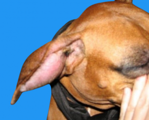 Hematoma on the Ear (Othematoma)